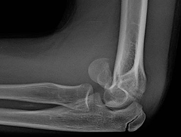
Capitellum Fractures
Capitellum fractures are a discrete subset of coronal plane partial articular injuries of the distal humerus, accounting for less than 1% of all elbow fractures. These are coronal shear fractures that may affect the capitellum alone, expand medially to include the trochlea, or occur in conjunction with lateral column osseous or ligamentous lesions.
For partial articular fractures of the distal humerus, several classifications have been devised, including those by Ring, Dubberley, and the AO/OTA. The Bryan and Morrey classification is the most widely used classification system. Type I fractures (Hahn-Steinthal) are complete capitellar fractures with little or no extension into the lateral trochlea, type II fractures (Kocher-Lorenz) are anterior osteochondral fractures with minimal subchondral bone, type III fractures are comminuted or compression fractures of the capitellum, and type IV fractures are capitellar fractures that extend medially to include mounds of bone.
Step by Step Procedure:
- Following general or regional anaesthesia, the damaged elbow is clinically evaluated for ligamentous stability under fluoroscopic supervision.
- A lateral skin incision at the elbow is positioned over the lateral epicondyle with the elbow flexed. From the anterior aspect of the lateral column to about 2cm distal to the radial head, this incision is made.
- The subcutaneous tissue layers are dissected and the lateral column is identified with the forearm in pronation to shift the posterior interosseous nerve away from the surgical field.
- The anterior capsule and the shared origin of the radial wrist extensors are lifted and moved anteriorly as a full-thickness flap from the lateral supracondylar ridge.
- Depending on the damage pattern, many deep intervals may be used distally. Kaplan (interval between the extensor digitorum communis and extensor carpi radialis brevis muscles), Kocher (interval between the aconeus muscle and the extensor carpi ulnaris muscle), and Hotchkiss (medial “over-the-top” approach between the flexor-pronator mass and the flexor carpi ulnaris muscle) are among them. To create a continuous anterior full-thickness soft-tissue flap, the distal gap is joined to the proximal exposure. We propose a more anteriorly based distal interval for capitellar fractures with or without proximal radius fractures to assist fracture reduction and fixation.
- Intracapsular retractors are put over the medial column and deep to the brachialis and anterior capsule. This makes the anterior articular fracture segments and the radial head/neck more visible.
- Elevation of the posterior inferior aspects of the capitellar-trochlear segment, with or without supplementary bone grafting, is required when the posterior inferior aspects of the capitellar-trochlear segment are impinged.
- With orthogonal fluoroscopy, provisional K-wire fixation is obtained and anatomic reduction is confirmed.
- When the articular fracture section has enough subchondral bone, buried cannulated variable pitch headless compression screws are placed in an anterior-to-posterior path over guidewires (terminally threaded Hebert screws or fully threaded mini-Acutrak headless screws). Two screws are put in a divergent pattern to limit rotation and are sufficiently apart from one another to prevent iatrogenic capitellum fracture.
- Suture fixation and bioabsorbable implants are additional options for Bryan and Morrey type II osteochondral fragment fractures where there is inadequate subchondral bone for compression screw internal fixation. When there is a pure cartilaginous fragment with no subchondral bone, excision is permissible, and the donor location can be repaired with chondroplasty.
- In more intricate fracture patterns, further fixation may be required. Mini-fragment screws, threaded k-wires, and bioabsorbable pins for small osteochondral capitellum and trochlea fragments. lateral column fixation with pelvic reconstruction, precontoured, or locking plates for posterolateral comminution and suture anchors or transosseous sutures passed through drill holes for lateral comminution.