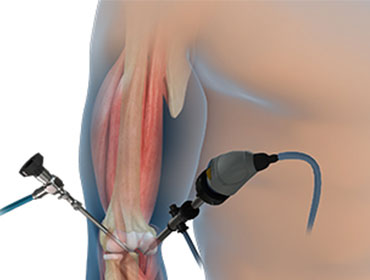
Elbow Arthroscopy
The term arthroscopy comes from Greek words that refer to the joint. In modern surgery, your surgeon uses a small camera and a video monitor to look inside your joint. This procedure allows your surgeon to make very small incisions, which are usually less than the size of a dime, to perform simple surgical procedures.
When elbow Arthroscopy Is Recommended?
If your condition does not respond to standard nonsurgical treatment, you may be considered for elbow arthroscopy. Nonsurgical treatment includes rest, physical therapy, and medications or injections that can reduce inflammation.
This procedure is performed to relieve pain and improve joint function.
Common arthroscopic procedures include:
- Treatment of tennis elbow (lateral epicondylitis)
- Removal of loose bodies (loose cartilage and bone fragments)
- Release of scar tissue to improve range of motion
- Treatment of osteoarthritis (wear and tear arthritis)
- Treatment of rheumatoid arthritis (inflammatory arthritis)
- Treatment of osteochondritis dissecans (activity related damage to the capitellum portion of the humerus seen in throwers or gymnasts)
- Treatment of fractures
Positioning
You'll almost certainly be given general anaesthesia as well as intravenous antibiotics once you're in the operation room. Antibiotics are commonly given prior to surgery to reduce the risk of infection.
After that, you'll be positioned such that the surgeon can easily move the arthroscope so that he or she can see the inside of your elbow clearly.
Lateral decubitus (side laying) and prone are the two most usual postures for elbow arthroscopy (lying on your stomach). After placement, extra care is given to protect and cushion your spine and other pressure points in your arms and legs.
Your upper arm is then wrapped in a tourniquet and placed in an arm holder to keep it in place during the treatment. To reduce swelling, apply a compressive dressing to your lower arm and hand. The surgical team will use antiseptic to clean your skin and sterile surgical drapes to wrap your shoulder and upper body.
Typically, surgeons draw lines on the elbow to highlight specific structures (such as the ulnar nerve and olecranon bone), as well as incision sites and arthroscope portals.
Procedure:
Your surgeon will first inject fluid into the elbow joint. The fluid allows your surgeon to see the structures of your elbow more clearly through the arthroscope's camera. The blood vessels and nerves that surround your elbow joint are less likely to be injured as a result of this. To insert the arthroscope and small instruments into the joint, your surgeon will make many small incisions. To maintain the view clean and control any bleeding, fluid flows through the arthroscope.
On the video screen, images from the arthroscope are shown, showing your surgeon the interior of your elbow and any concerns. Before commencing any specific therapies, your surgeon will assess the joint.
If necessary, the entire joint will be examined, which could necessitate five or seven extremely small arthroscopic incisions. Other small instruments will be inserted by the surgeon through separate incisions to help with the surgery. Shaving, cutting, gripping, suture passing, and knot tying are all done with specialised devices. Special devices are frequently employed to anchor stitches in bone.
At the conclusion of the surgery, the arthroscopy incisions are usually stitched or covered with skin tapes. The elbow is dressed with an absorbent dressing. Your surgeon will apply either an additional soft dressing that allows movement or a plaster splint that restricts movement and protects the elbow, depending on the treatment.