Floating Shoulder
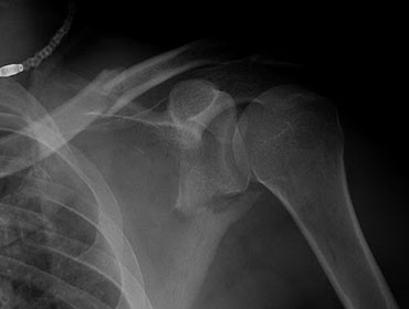
Ipsilateral fractures of the midshaft of the clavicle and the neck of the glenoid are known as the floating shoulder. Without a detailed understanding of the complicated anatomy of the shoulder girdle, this uncommon ailment can be difficult to manage. For all of these injuries, surgical intervention should be considered. While non-operative care of mildly displaced fractures can yield satisfactory outcomes, displacement at one or both sites is best addressed with surgical reduction and fixation.
Surgical Procedure:
Wide access to the entire shoulder girdle is required for operations on the floating shoulder. The patient is positioned in the lateral decubitis or ‘deck chair' position. The scapula and clavicle must be exposed to the full extent possible. The entire upper limb, including the shoulder girdle, is prepped and draped. If the surgeon prefers, a staged procedure with separate postures, sterile preparations, and exposures can be undertaken.
The order in which the bones are fixed is controversial and at the surgeon's discretion. Initial clavicle fixation may allow for indirect glenoid segment reduction, obviating the need for a subsequent surgery. However, if substantial displacement persists, the glenoid fracture must be addressed as well. Fixation of the displaced glenoid segment, on the other hand, may be judged more significant and performed first, followed by open reduction and internal fixation of the clavicle fracture, if necessary.
An incision is made directly above the subcutaneous border of the clavicle. The neighbouring neurovascular structures must be identified and protected. A conventional plate and screws are used to decrease and stabilise the fracture site, which is exposed subperiosteally both proximally and distally. In some cases, such as severe comminution, osteoporotic bone, or surgeon preference, intramedullary devices, precontoured plates, and locking plates may be recommended.
Backwards, the glenoid neck is approached. The posterior deltoid is either divided along its fibres or disconnected and retracted distally. To reveal the posteroinferior glenoid neck and lateral edge of the scapula, the space between infraspinatus and teres minor is developed. To manage the free glenoid fragment, a superior approach can be introduced. Temporary fixation can be performed by inserting K-wires through the glenoid fragment and into neighbouring bony structures once a suitable reduction has been achieved (for example, through the glenoid fragment and into the scapular body, or through the acromial process into the glenoid fragment). A contoured 3.5 mm reconstruction plate is usually used to establish definitive fixation along the lateral border of the scapula and the posterior portion of the glenoid process. K-wires or lag screws can be used to offer additional support. The temporary fixation K-wires can be left in place or utilised to install 3.5 mm cannulated lag screws.
In rare situations, comminution of the scapular body and spine is severe enough, or the glenoid fragment is small enough, that plate fixation is impossible. In these cases, K-wires or lagged screws may be the sole way to secure the decreased glenoid fragment to the nearby undamaged bony structures, such as the acromial process or the distal clavicle.